Stability of proximal mandibular anatomical structures following bilateral sagittal split osteotomy
Authors
- Holte MB, Nielsen TW, Cattaneo PM, Pinholt EM

Abstract
Objectives
The aim of this study was to investigate the stability of the proximal mandibular reference structures that have been proposed in the literature for superimposition.
Methods
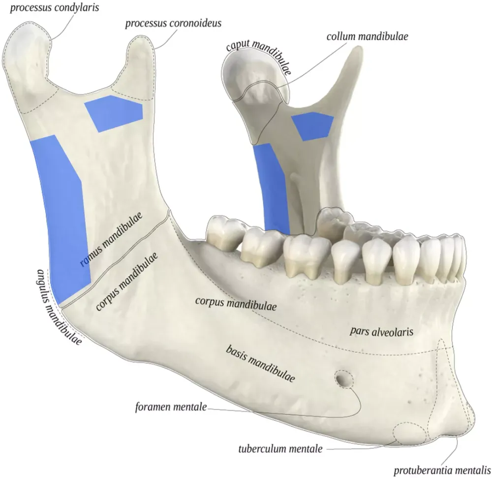
Forty proximal mandibular segments of 20 patients who underwent bilateral sagittal split osteotomy (BSSO) for advancement were reconstructed from a pair of pre- and postoperative (2 years) cone beam computed tomography scans, and spatially divided into the mandibular condyle, the coronoid process, and 20 mandibular ramus regions. To assess the stability of the anatomical regions, the volumetric and surface discrepancy between the superimposed pre- and postoperative regions were calculated. One-sample t-tests were applied to analyse the statistical stability of the individual regions.
Results
Two statistically stable (P < 0.05) structures in the proximal segment of the mandible following BSSO were identified: (1) the posterior part of the mandibular ramus above the gonial angle and below the condylar neck, and (2) the sub-coronoid area below the coronoid process/mandibular notch. Using these stable structures for superimposition resulted in an assessment discrepancy in the condylar displacement of up to 1.1 mm and in the volumetric change of up to 2.8%.
Conclusion
Hence, it is suggested that these two identified stable structures are used as reference areas when assessing condylar displacement and change using superimposition.